
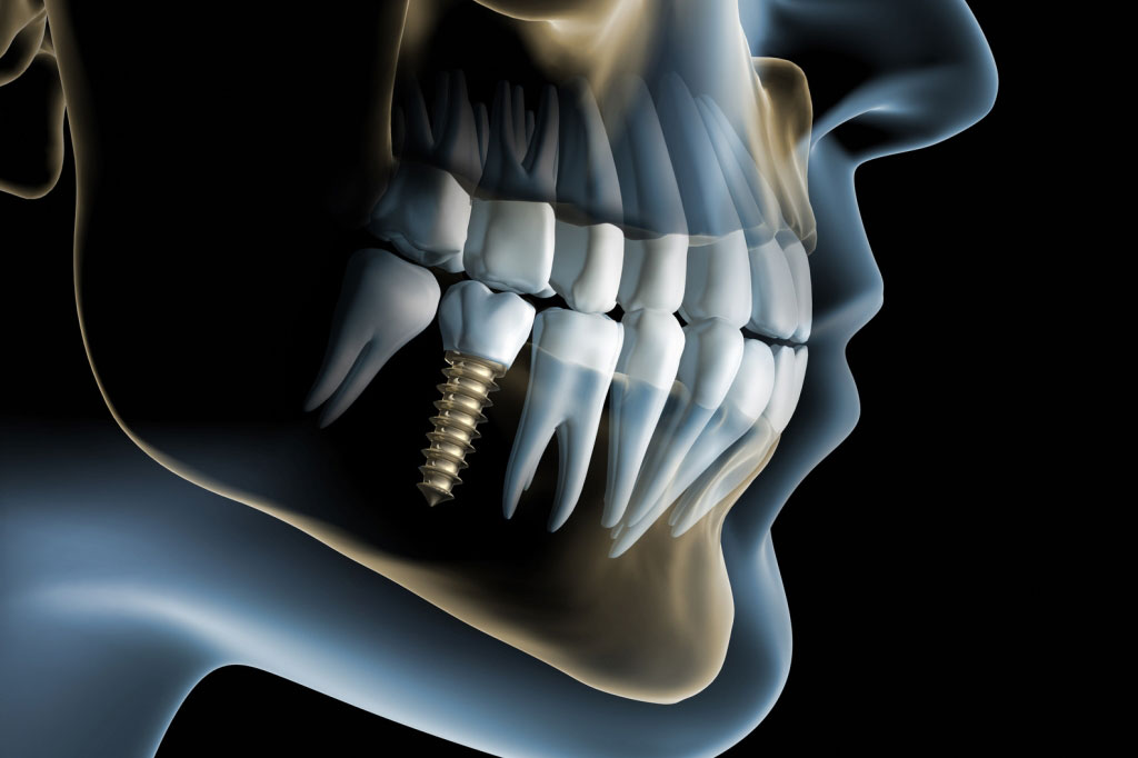
A dental implant is specially designed structure placed in the jawbone whose role is to fully simulate the function of…
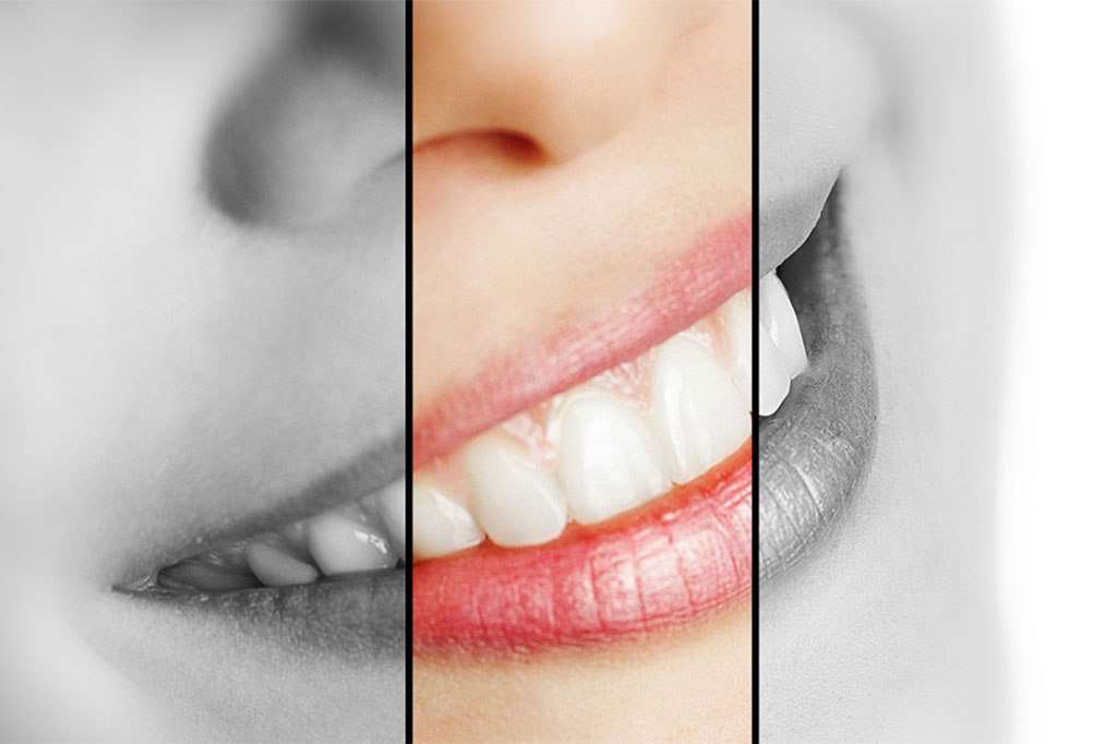
Good looks has become an imperative of modern man. The teeth and a beautiful smile are definitely one of the…
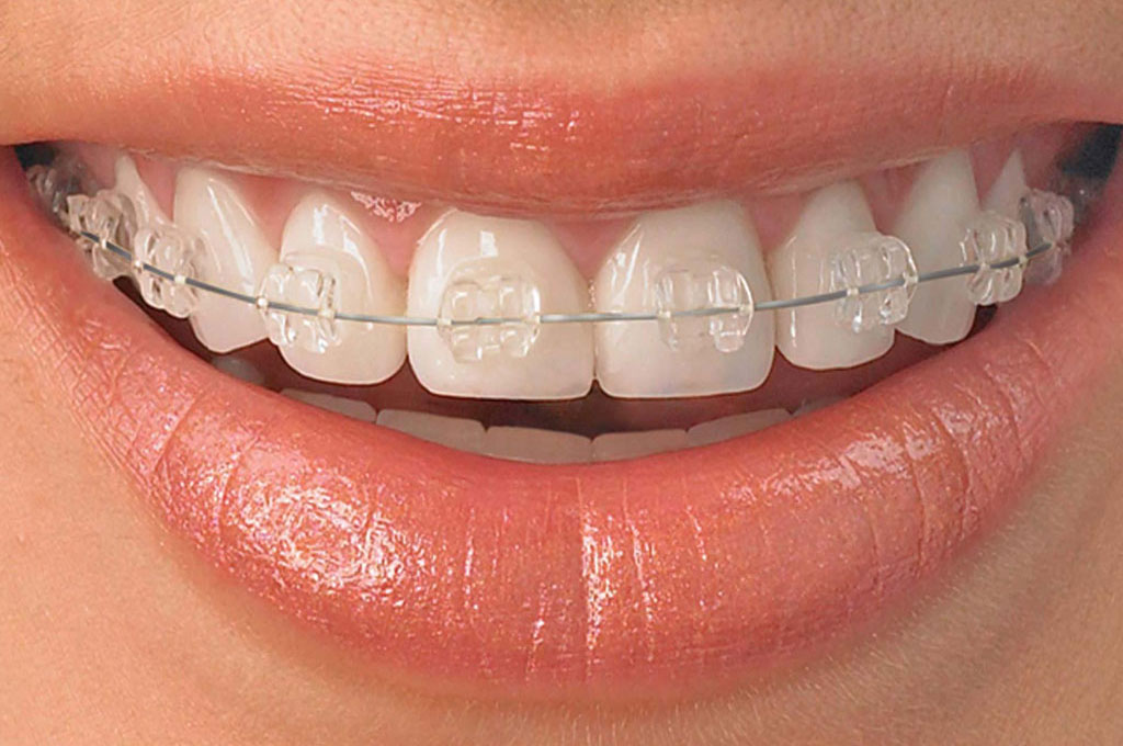
Orthodontics (orthodontics) is a branch of dentistry that deals with the prevention, diagnosis and correction of congenital and acquired tooth

Periodontics is the area of dentistry that deals with prevention, diagnosis and treatment of gum disease and tooth supportive tissues.
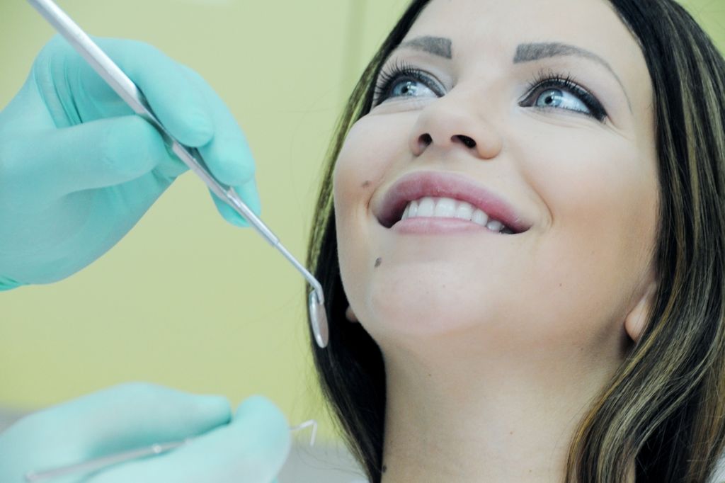
Oral surgery is a specialized field of dentistry that deals with the diagnosis and treatment of many diseases and conditions…
Teeth whitening are one of the basic methods of cosmetic dentistry. It is based on the oxido -reductive processes in…
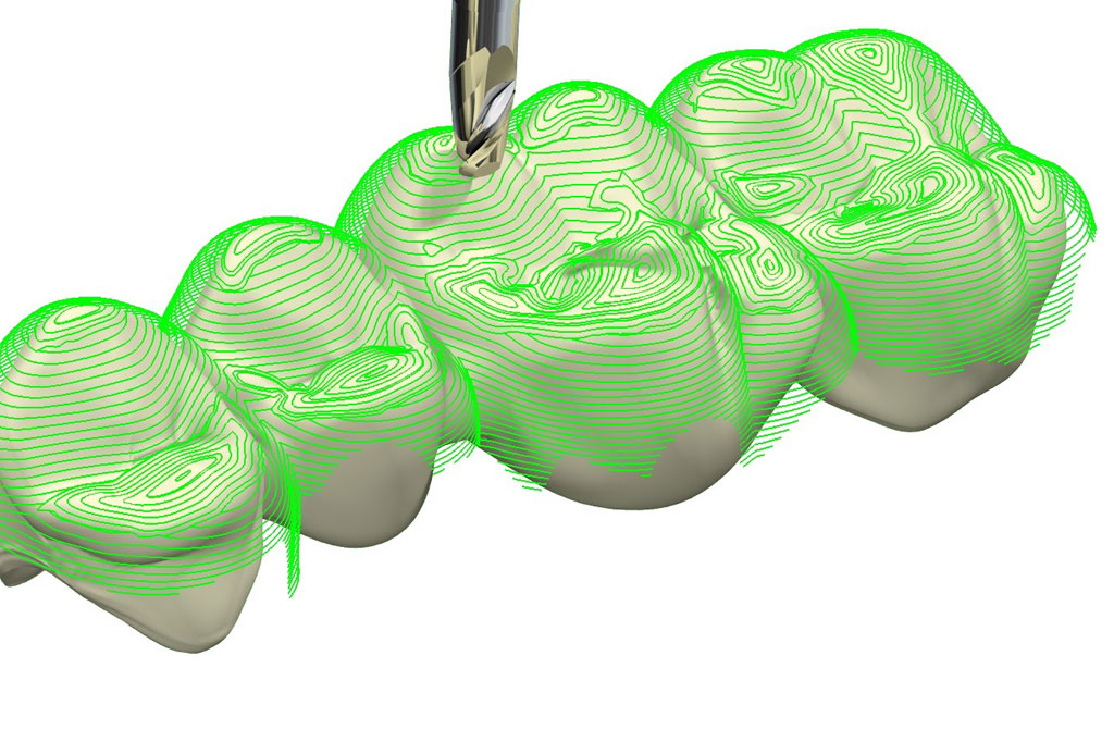
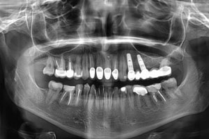
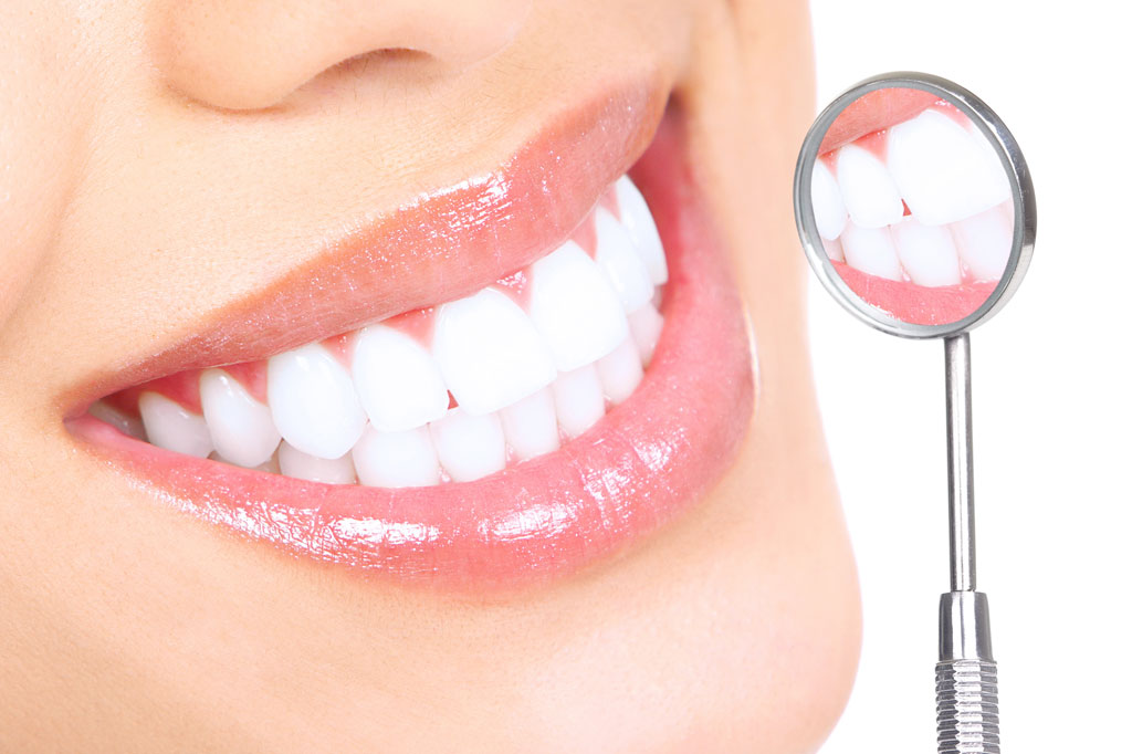
Prosthodontics is the field of dentistry that deals with the reconstruction of damaged teeth, replacement of lost tooth or group…

Children are born with all the primary teeth that are not visible because they are underneath the gums. Eruption of…


